


Cervical osteochondrosis of the neck is a common pathology that is accompanied by unpleasant symptoms. The disease is characterized by degenerative-dystrophic processes in the spine. They are caused by incorrect posture, poor posture and poorly active lifestyle. In order to reduce the symptoms of the pathology, you should consult a doctor in a timely manner.
What is cervical osteochondrosis?
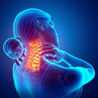
This term is understood as a progressive degenerative-dystrophic lesion of the intervertebral discs located in the cervical spine. As a result, the vertebral bodies are deformed and exhausted. This leads to an impairment of the blood circulation and the nerve conduction in the neck.
The disease can be isolated or combined with damage to other parts of the spine - lumbar or thoracic. According to ICD-10, cervical osteochondrosis of the spine is coded under the code M42.
Possible complications of the disease
Many people are interested in the risk of cervical osteochondrosis. Without timely and adequate therapy, the pathology can lead to negative health consequences. These include the following:
- protrusion of the intervertebral discs and herniation;
- Intervertebral disc rupture with compression of nerves and blood vessels - if the spinal cord is pinched, there is a risk of death;
- radiculopathy;
- the appearance of osteophytes;
- Paresis and paralysis.
The main symptoms and signs of cervical osteochondrosis
The disease develops gradually and is initially asymptomatic. Therefore, the diagnosis is often made in advanced cases. The main symptoms of the pathology are the following:
- Pain in the neck and occiput, worsened by physical exertion or coughing.
- Grinding when moving the head.
- Loss of sensitivity in the hands, burning sensation in the shoulder blades.
- Headache that is localized in the back of the head and spreading to the temples.
- General weakness, increased tiredness.
- Decreased visual acuity.
- tinnitus.
- hearing loss.
- Increased heart rate.
Causes of cervical osteochondrosis
The first signs of the disease usually appear after 35 years. However, in recent years, the pathology began to develop at a younger age, 18-30 years old. Most often, the problem is faced by people who have to be in one position for a long time.
The main causes of cervical osteochondrosis are:
- hereditary tendency;
- violation of metabolic processes;
- infectious diseases, poisoning of the body;
- disorders in nutrition - lack of fluids, vitamins, microelements;
- overweight;
- traumatic injuries to the spine;
- bad posture;
- instability of the spine;
- insufficiently active lifestyle;
- flat feet;
- Influence of unfavorable environmental factors;
- frequent changes in posture;
- being in an uncomfortable position for a long time;
- excessive physical activity;
- hypothermia;
- stressful situations;
- with the wrong sleeping pillows.
What are the different degrees of illness?
The disease develops gradually. There are 4 degrees of cervical osteochondrosis, each with specific characteristics:
- The first is accompanied by the appearance of cracks in the intervertebral discs. This process is accompanied by mild pain and stiffness in movements. The pathology has a wavy course. With the deterioration in the immune system or an increase in the load, osteochondrosis worsens. If you do not take action in a timely manner, there is a risk that the abnormal process will worsen.
- Second - at this stage, the destruction of the intervertebral discs continues and their protrusion is observed. This process is accompanied by pinching the nerve endings. A person has constant pain that increases with movement. At this stage, the ability to work decreases, numbness in the hands appears.
- The third is accompanied by the appearance of an intervertebral hernia. In such a situation, muscle tissues and nerve endings are involved in the pathological process. As a result, there is pain in the neck and the back of the head, a feeling of weakness in the hands. With vascular lesions, there is a risk of decreased visual acuity, dizziness, and tinnitus. Sometimes the disease leads to fainting.
- Fourth - this stage is accompanied by bone growth. As a result, the pressure on the nerve endings increases. In this form of osteochondrosis, the mobility of the neck decreases and the spine becomes less flexible. As a result, a person cannot make simple head movements.
Why should you consult a doctor immediately?
If symptoms of osteochondrosis appear, you should immediately consult a doctor - a neurologist or orthopedic surgeon. Otherwise, the pathology will lead to dangerous health consequences.
First of all, the doctor should assess the physical activity and the intensity of the neck pain. The specialist is also interested in loss of sensitivity and other disorders.
Based on the results of the preliminary examination, additional procedures will be prescribed. First, a radiography is done. It is carried out in several projections. If an inguinal hernia is suspected, CT or magnetic resonance imaging may be required. With a violation of blood flow, rheoencephalography and examination of the fundus are required.
Treatment is prescribed based on the results of the diagnostic examination. With the development of cervical osteochondrosis, the following categories of drugs are most often used:
- Analgesics - help manage pain.
- Non-steroidal anti-inflammatory drugs - eliminate inflammation and manage swelling.
- Antispasmodics - help with muscle cramps.
- preparations to improve blood circulation.
- Chondroprotectors - help to restore the structure of the intervertebral discs.
- B vitamins - improve the functioning of the nervous tissue.
In addition to drug therapy, other methods are prescribed. This includes massage, remedial gymnastics, physiotherapy. The use of osteopathy is very effective. In this case, a mild effect is exerted on the affected muscles and vertebrae. In some cases, the doctor is advised to wear a special orthopedic device - the Shants collar.
Manual therapy is considered an effective method of treating pathology. Your methods are selected individually. The procedure consists in a point effect on the musculoskeletal elements. Thanks to this, it is possible to activate blood flow, improve lymphatic movement and normalize metabolic processes. Manual therapy improves the mobility of the musculoskeletal system, strengthens the immune system and prevents complications from osteochondrosis.
Spinal traction is widely used. Special equipment is used for stretching. The procedure helps increase the distance between the vertebrae to normal size and cope with disruptions in the structure of the spine.
If acute cervical osteochondrosis is observed and intervertebral hernias appear, leading to a decrease in sensitivity and impaired blood flow, surgical intervention is required.
The duration of treatment depends on the severity of the osteochondrosis. Most often, therapy is carried out in long courses. Definitely, in order to improve your condition, you should make adjustments to your lifestyle. To do this, you need to eat right, give up bad habits and exercise.
Prevention of osteochondrosis of the neck
To prevent the occurrence of cervical osteochondrosis, you must adhere to certain recommendations:
- timely eliminates the curvature of the spine;
- do exercise to build a muscle corset.
- eat foods that provide the body with calcium and magnesium;
- normalizes body weight;
- When using a computer, follow the recommendations of doctors.
Cervical osteochondrosis is a serious pathology that leads to negative health consequences. To cope with the violation, it is necessary to make the correct diagnosis in a timely manner. Therefore, discomfort in the neck area should be a reason to consult a doctor.
How is the treatment going?
Doctor's advice: anamnesis, myofascial diagnostics, functional diagnostics.
How are you?
Collect anamnesis - analyze illness, identify limitations and contraindications, explain the principles of kinesitherapy and the characteristics of the recovery phase.
Myofascial diagnostics is a method of manual diagnostics in which the doctor assesses the range of motion of the joints, identifies painful seals, edema, hypo- or hypertonicity of the muscles and other changes.
Functional diagnostics (carried out in the rehabilitation room) - The doctor explains how certain exercises are carried out on the devices and observes: how the patient performs them, which range of motion he can work with, which movements cause pain, whichThe patient's weight can work on how the cardiovascular system reacts. Problem areas are identified. The data is entered into the card. The focus is.
Based on the results of the initial examination by a doctor and the functional diagnosis, a preliminary individual treatment program is drawn up.
It is desirable to have the following with you:
- for pain in the spine - MRI or CT scan (magnetic resonance imaging or computed tomography) of the problem area;
- for joint pain - X-rays;
- in the case of comorbidities - extracts from the medical history or an outpatient card;
- comfortable (sports) clothing and shoes
Start the lesson with a teacher
At the beginning of the treatment cycle, the doctor works with the patient to create a treatment plan that includes the date and time of the treatment session and follow-up examinations with the doctor (usually 2-3 times a week).
The basis of the treatment process is treatment sessions in the rehabilitation room with exercise equipment and sessions in the gym.
With rehabilitation simulators you can precisely dose the load on individual muscle groups and thus achieve an appropriate physical effect. The treatment program is drawn up by the doctor individually for each patient, taking into account the characteristics of the organism. The inspection is carried out by qualified instructors. At all stages of recovery, it is important to observe the technique of correct movement and breathing, know your weight standards when working on simulators, adhere to the prescribed treatment regimen and follow the recommendations of specialists.
Joint gymnastics sessions help restore visual coordination, improve joint mobility and the elasticity (flexibility) of the spine, and are an excellent prevention system for independent use.
Each treatment cycle - 12 sessions. Each lesson is supervised by a teacher. The duration of a treatment is 40 minutes to 1. 5 hours. The instructor creates a program that takes into account the comorbidities and condition of the patient on the day of training, teaches the technique of performing exercises and monitors the correctness of execution. A second consultation with a doctor is carried out every 6th hour, changes and additions to the program are made depending on the dynamics.
How many loops are required?
This is individual for each person and depends on the progression of the disease.
Important to know:
- How long have you had this problem (stage of the disease)?
- how your body is prepared for physical activity (do gymnastics, any kind of sport);
- what result you want to get.
If the disease is in its early stages and the body is prepared, one cycle of treatment is sufficient. (Example - young people aged 20 to 30 who play sports. We focus their attention on technique of doing exercises, breathing, stretching, and eliminating "wrong" exercises that are harmful to problem areas. Such patients are trainedand maintain the ability to take care of your body », Receive recommendations in the event of an exacerbation and keep doing it yourself)
Every organism is individual and the program for each patient is individual.